Last year Olympus launched the first-ever Olympus Image of the Year Award – a global photography competition for scientific images which recognises their simultaneous artistic value.
The winners of the 2019 competition have just been announced and the mindblowing images offer a rare insight into the ethereal world of the all-present microcosmos. Check them out below.

The winners of the inaugural Olympus Image of the Year Award have been unveiled and they offer a breathtaking peek into the world of the microcosmos.
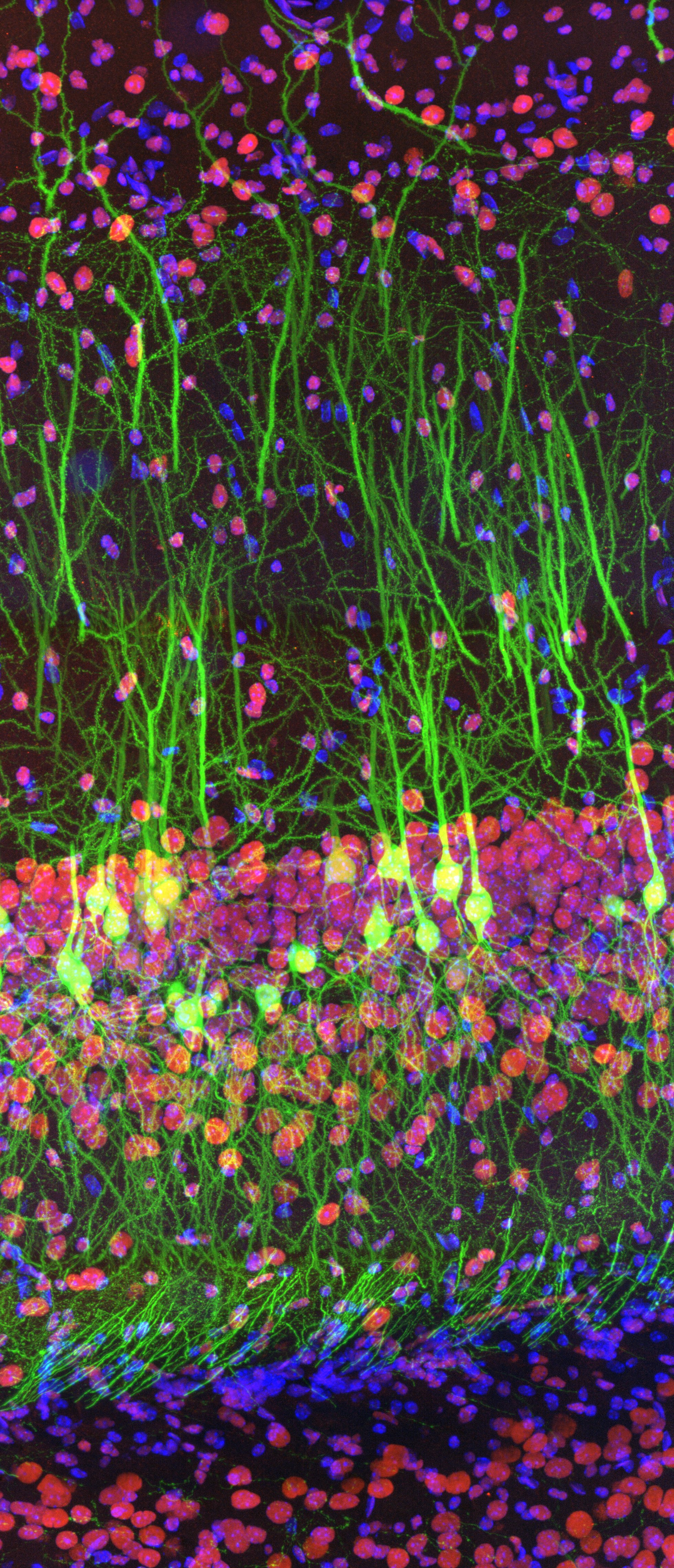
The competition has one overall winner and three regional winners – from the Americas; Europe, the Middle East and Africa; and Asia-Pacific respectively. The 2019 overall winner was a photograph of a mouse’s hippocampus (the part of the brain involved with memories) from Ainara Pintor, a graduate researcher in Spain.
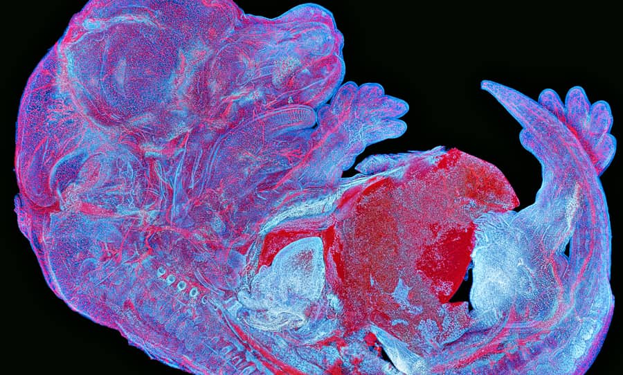
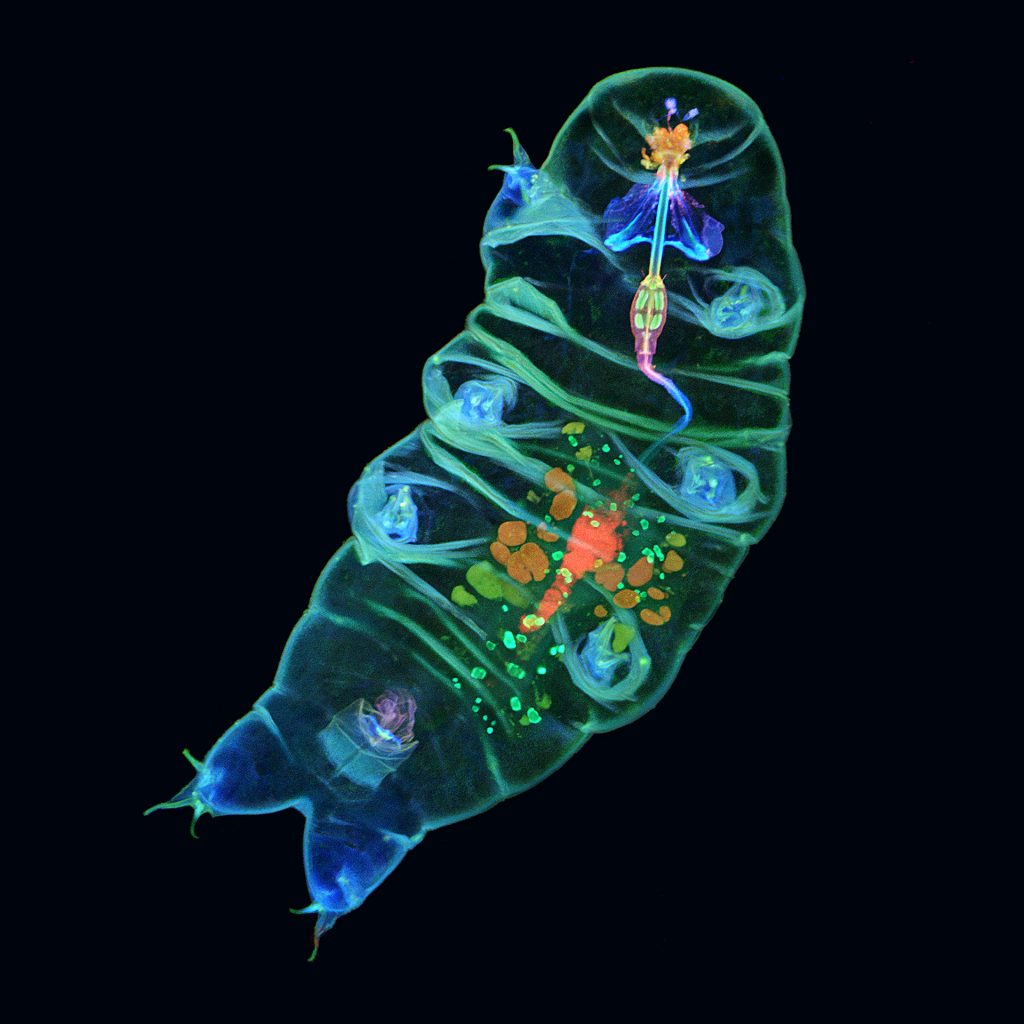
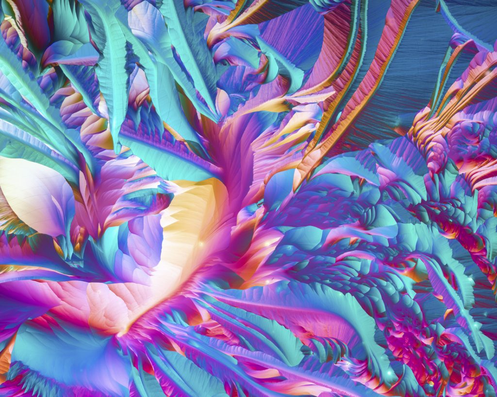
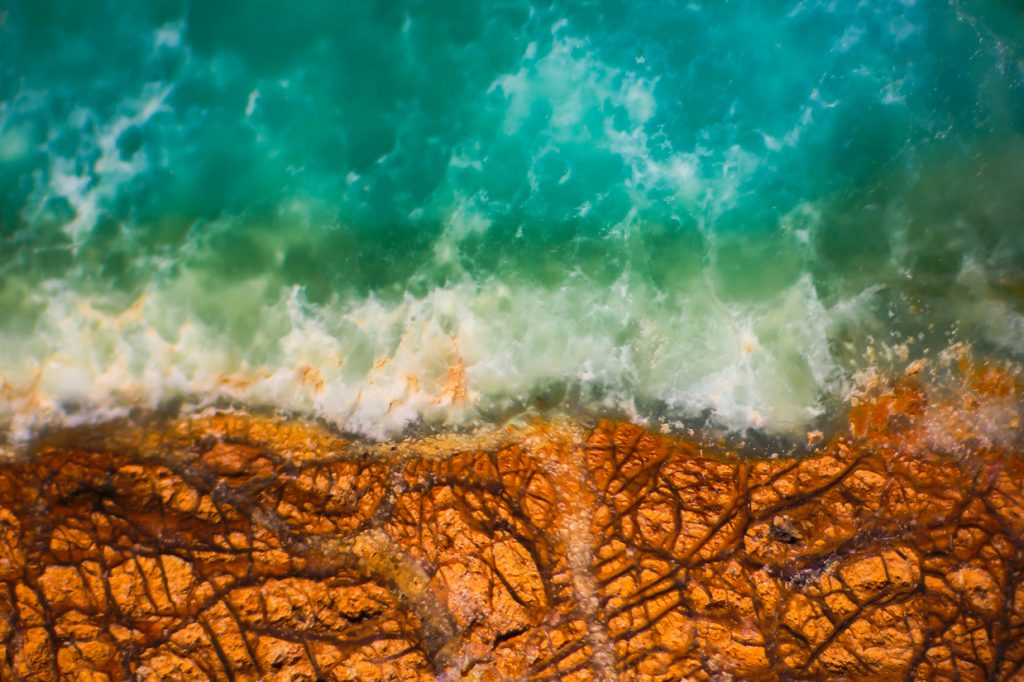
Some of the other entries include the autofluorescence of a mouse embryo, the inside of a tardigrade (a micro-animal known as a moss-piglet), and a magnified prase opal, which bears an uncanny resemblance to an ocean coastline.
Check out the winners and some honourable mentions below.
Next Up: Check out these mind-blowing photos from The Perfect Moment Contest
The overall winner, taken by Ainara Pintor (Spain):
The regional winner for the Americas, taken by Tagide deCarvalho (U.S.A.):
The regional winner for Europe, the Middle East and Africa, taken by Alan Prescott (U.K.):
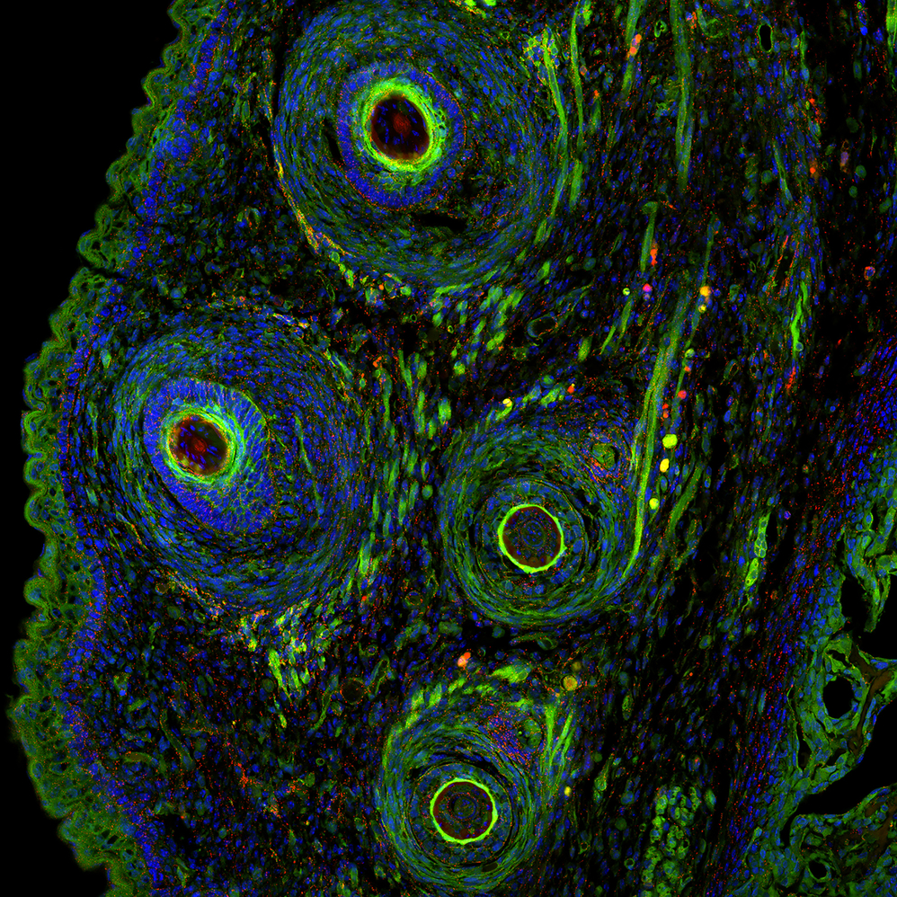
The regional winner for the Asia-Pacific, taken by Howard Vindin (Australia):

Honourable mention: Ming-Der Lin (Taiwan):

Honourable mention: Nat Prunet (U.S.A.):

Honourable mention: Justin Zoll (U.S.A.):
Honourable mention: Nathan Renfro (U.S.A.):
To check out more entries, head over to the Olympus Image of the Year Award website.